¹ Bureau of Labor Statistics, Occupational Outlook Handbook, Optometrists, 2023–24 edition.
🔬
Built by a practicing optometrist
OD — Doctor of OptometryActive clinical practiceOCT/OCTA hands-onVendor-neutral
SlabED was created by an optometrist who reads OCT and OCTA scans in daily practice — not a vendor, not a CE company, not an AI. The curriculum comes from real clinical experience: the interpretation questions that come up in exam lanes, the pathology patterns that change management, and the en face knowledge that optometry programs skip entirely.
Vendor-neutral by design. No equipment partnerships. No sales agenda. Just the science.
What's included
19 lessons. 3 tiers. Every clinical scenario that matters.
From retinal layer anatomy to full OCTA interpretation — structured, clinically rigorous, and completely free. No credit card. No paywall. No trial period. The full curriculum, open the moment you sign up.
AMD Drusen Mapping
Distinguish hard drusen, cuticular drusen, and drusenoid PED on en face. Know when fellow-eye risk changes your monitoring interval.
Diabetic Macular Edema
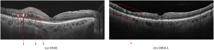
Identify DRIL, intraretinal cysts, and hard exudates — the OCT features that predict visual outcome and trigger referral.
Glaucoma Progression
Use RNFL and GCC analysis alongside en face to catch structural loss before your patient notices. Correlate disc changes with OCT findings.
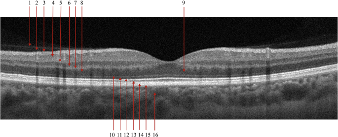
Before you can identify pathology, you need to own the normal. This lesson walks through all ten retinal layers visible on OCT — from the internal limiting membrane (ILM) at the top to the choroid below — with the specific hyperreflective and hyporeflective bands that define each one.
EZ band — the ellipsoid zone (bright hyperreflective line) is your photoreceptor health marker. Disruption here means outer retinal damage.
RPE — the retinal pigment epithelium sits directly above Bruch's membrane. Drusen, PED, and CNV all distort this layer first.
RNFL — nerve fiber layer thinning on OCT is the earliest structural sign of glaucoma, often visible before field loss.
Choroidal vessels — irregular hyporeflective spaces below the RPE. Pachychoroid pattern and lesions originate here.
Real Clinical Scans
This is what you'll learn to read.
Real OCT B-scans from peer-reviewed open-access research — the same pathology patterns covered in every SlabED module.
Normal Macula · B-Scan
Normal macular B-scan Image loads from Springer CDN
Ten retinal layers from ILM (top) to choroid (bottom). EZ band (ellipsoid zone) and RPE appear as bright hyperreflective bands above the dark choroid.
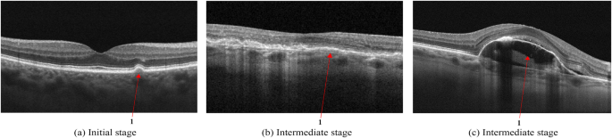
AMD Drusen · B-Scan
AMD drusen OCT examples Image loads from Springer CDN
AMD drusen spectrum: hard drusen (small focal RPE deposits), cuticular drusen (sawtooth pattern), and drusenoid PED (broad flat RPE elevation).
Diabetic Macular Edema · B-Scan
DME OCT example Image loads from Springer CDN
Diabetic macular edema: retinal thickening, intraretinal cysts, hard exudates (hyperreflective foci), and DRIL — the strongest OCT predictor of poor VA.
You learned B-scan in school. Nobody taught you en face.
En face imaging reveals pathology that cross-sectional B-scans miss entirely. But optometry programs barely cover it, CE courses skip it, and equipment reps assume you already know it. The result: practices running $40K machines at half their diagnostic potential.
?
Education gap
No structured curriculum exists for en face interpretation. ODs are guessing or avoiding the modality entirely.
$
Blocked purchases
A $10K–$15K OCTA upgrade unlocks capabilities most ODs aren't trained to use. Practices stall on the investment — not from cost, but from uncertainty about return.
!
Vendor lock-in
Equipment reps teach their platform, not the science. You need vendor-neutral education to make the right purchase.
Learning Pathway
Three tiers. One destination: clinical confidence.
Start where you are. Every optometrist follows a structured path from their current skill level to full OCTA fluency.
10 clinical scenarios where en face changes management
Artifacts, segmentation errors, and documentation
03
OCTA Integration
For ODs ready to add angiography
OCT-A flow signal and vascular mapping
Four-modality analysis: B-scan + en face + OCTA + structural
Equipment comparison: vendor-neutral buying guide
ROI modeling for your practice
CPT 92137 billing: when to use it, ~$57 reimbursement, what to document
Curriculum Highlights
Built from real clinical cases
Every module maps imaging findings to management decisions. Not theory for theory's sake.
Four-Modality Imaging
Learn to synthesize B-scan, en face, OCT-A, and structural en face findings for AMD, diabetic retinopathy, RVO, CSC, ERM, macular holes, and choroidal lesions.
Layer-by-Layer En Face
ILM, superficial capillary plexus, deep capillary plexus, outer retina, RPE/Bruch's, choriocapillaris, and choroid. Know which layer matters for which pathology.
Management-Changing Scenarios
10 clinical scenarios where en face imaging directly changes your treatment plan. CNV differentiation, macular ischemia detection, geographic atrophy monitoring, and more.
Equipment Decision Framework
Vendor-neutral comparison of Zeiss, Topcon, Heidelberg, and Optovue platforms. Understand what you need before you spend $60K.
Getting Paid for OCTA: CPT 92137 & Practice Economics
The 2026 OCTA-specific billing code — ~$56.93 nationally, higher than any standard OCT code. Learn when you can bill it, what documentation is required, which codes it cannot be combined with, and how the 2026 Stark Law update affects your practice. The module ODs need before they adopt OCTA.
Stop guessing at scans. Start reading them with confidence.
SlabED is the only structured education platform dedicated to en face OCT imaging and OCTA for optometrists. Weekly lessons, clinical cases, and a clear pathway from wherever you are to wherever you need to be.
Free
No subscription. No credit card. All lessons included.
Each issue: one en face scan breakdown, one clinical interpretation tip, one equipment or billing insight. No fluff — just the kind of case discussion that makes the next scan easier to read.